Understanding the cardiac conduction system

The cardiac conduction system, responsible for controlling the coordinated contraction of the heart to maximise pumping efficiency, comprises many complex structures. Its function is critical to the healthy working of the heart, and dysfunction of the cardiac conduction system may underpin many cardiac conditions.

In combination with experimental collaborators in Manchester, Liverpool and Auckland, we are exploring methods to develop computational models of the intact cardiac conduction system that can be used to study its (dys)regulation in disease.


Concept: The Action Potential

Cardiac cells are encased by a cell membrane. Differences in the concentration of charged particles (called ions) inside and outside the cell result in a membrane potential - a difference in electric potential across the membrane. The Action Potential refers to the change in this membrane potential during excitation, illustrated below.

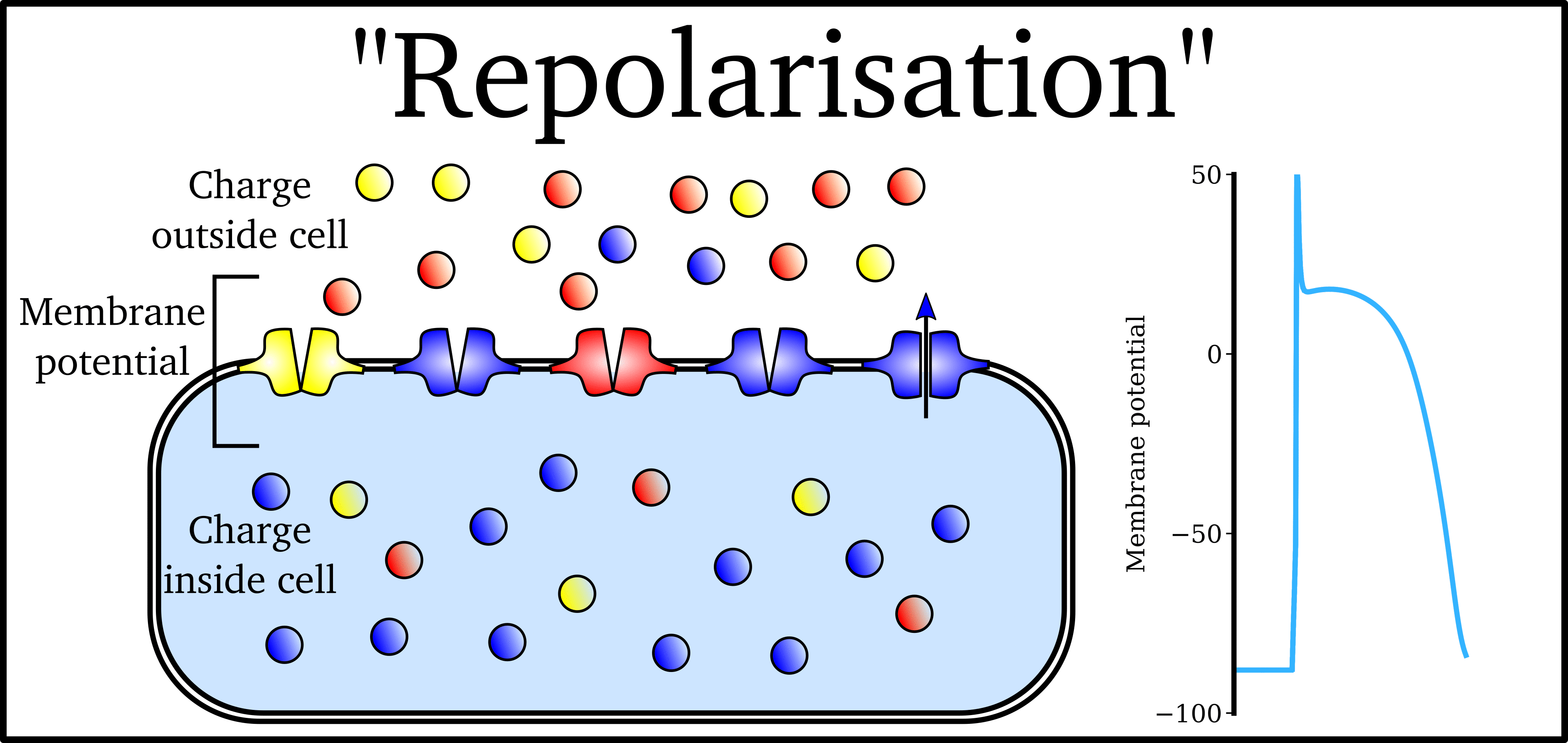
Animation: The Action Potential. Left panel shows a schematic of a cell with different types of ion and the channels which allow them to pass (coloured circles and channels), and the right panel shows the membrane potential at different stages of the Action Potential.

Ions can cross the cell membrane through specialised proteins (called ion channels), which can open or close to allow or prevent the passage of ions.

As ions flow through these ion channels, the movement of charge creates ionic currents which can change the membrane potential. Because the opening and closing of ion channels can also be controlled by the membrane potential, the currents can be coordinated to create the shape of the Action Potential.

There are multiple types of ion channel, each associated with specific ions (sodium, potassium, calcium and others).

"Inward" currents are those where positive charge enters the cell (yellow and red in the animation) - they make the membrane potential more positive, which we call “depolarisation”: Larger inward currents generally mean greater excitability and longer durations of the Action Potential.

"Outward" currents are those where positive charge leaves the cell (blue in the animation) – they make the membrane potential more negative, which we call "repolarisation": Larger outward currents generally mean lower excitability and shorter duration.

The balance of multiple different currents, controlled by the expression (amount of) each type of ion channel, can therefore control Action Potentials with different shapes and properties to match the varying requirements of cardiac function in different animals and under different conditions (e.g. rest versus exercise).


Concept: The Cardiac Conduction System

This refers to the specialised system within the heart which ensures it contracts in a coordinated manner to give the most efficient pumping function. By initiating the heartbeat in a specialised pacemaker region which spontaneously generates Action Potentials, and then conducting the excitation along specialised networks which can either delay or accelerate the propagation, a uniform and controlled contraction can be maintained.

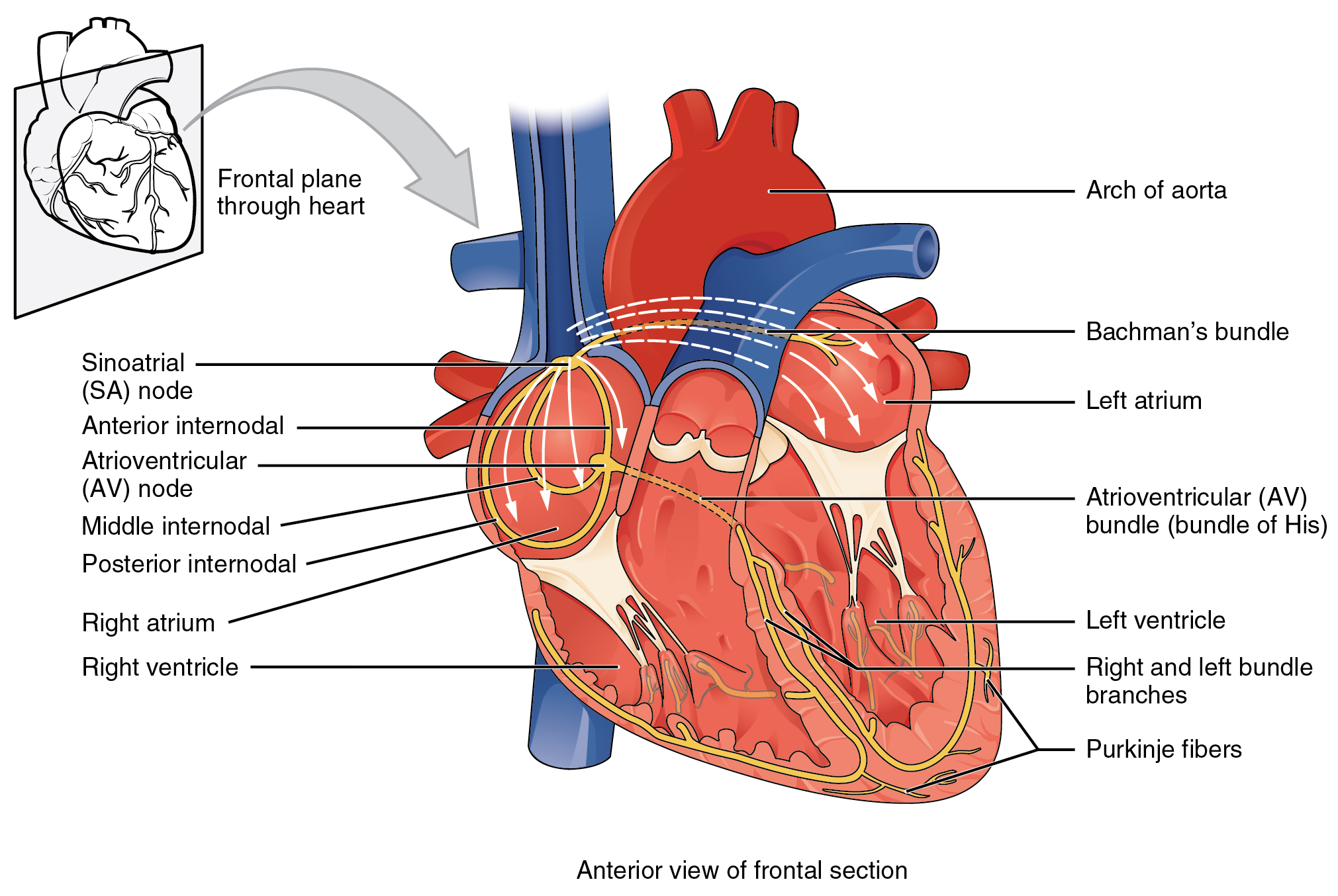
Illustration of the cardiac conduction system. Source: here (public domain) Attribution: OpenStax College [CC BY 3.0 (https://creativecommons.org/licenses/by/3.0)]

On each heartbeat, the following process occurs:
  1. The electrical excitation is initiated in the "sino-atrial node", the natural pacemaker, located near the top of the right atrium.
  2. The excitation is conducted along preferential pathways in the atria (the upper chambers of the heart) and down to the "atrio-ventricular node", the only electrical connection between the atria and ventricles .
  3. As excitation spreads throughout the atria, they contract and the ventricles fill with blood.
  4. Conduction is delayed in the atrio-ventricular node to allow time for the ventricles to fill.
  5. After this delay, excitation is conducted rapidly to the apex (the bottom) of the ventricles, through the bundle of His, before quickly spreading through the Purkinje Fibres throughout the ventricle. This supports near-simultaneous activation of the entire ventricular mass.
  6. The heart then undergoes repolarisation (returning to the non-excited state), allowing the atria and ventricles to fill in preparation for the next beat.


Researching the Cardiac Conduction System

Constructing a structure-based model of activation in the human CCS

In this publication, the team in Manchester demonstrate the use of contrast enhanced imaging techniques to segment and reconstruct the cardiac conduction system in multiple human heart samples.

They were able to identify and segment all of the core components of the cardiac conduction system, enabling analysis of its structural features in different datasets.

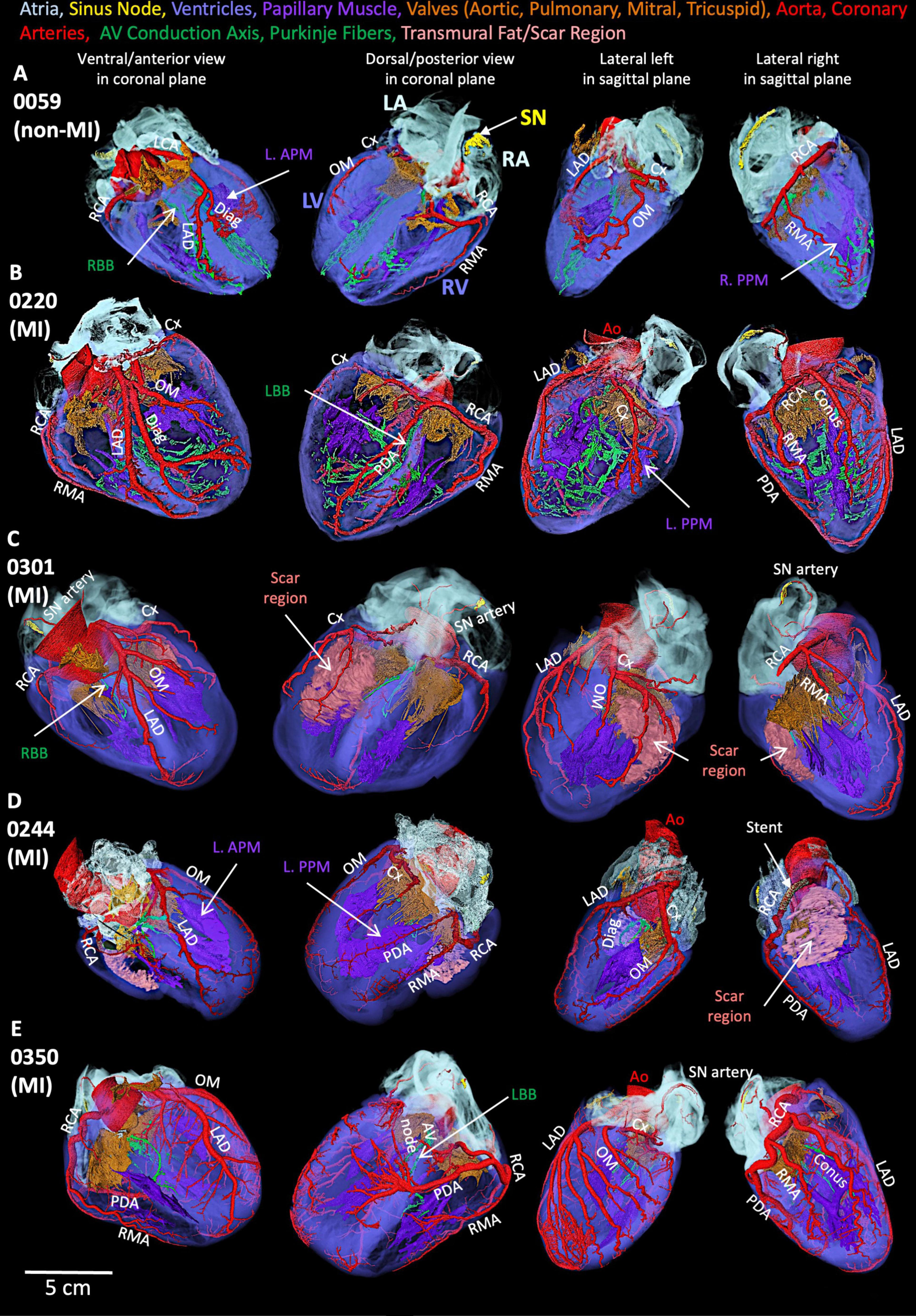
Figure: Segmented cardiac conduction system in five human heart smaples.

These data enabled the construction of a computational model that included the whole, intact system, able to reproduce the activation sequence in the human heart. This simple model demonstrates the value of the structural data, and we are currently developing this model further to include full details of electrophysiology and pacemaking in the different regions. Watch this space!

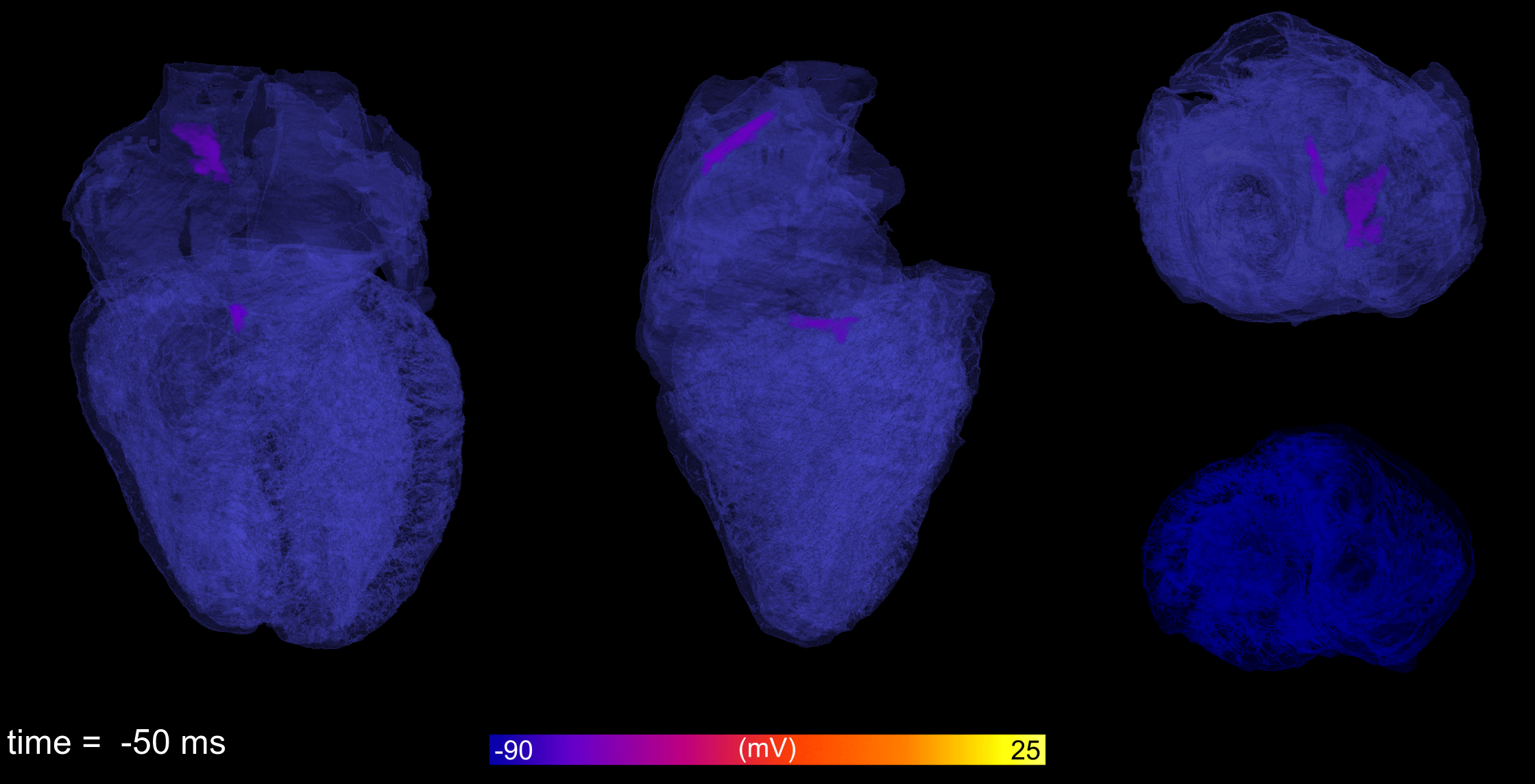
Animation: Simulation of the electrical activation sequence in a 3D model that includes the fully reconstructed cardiac conduction system.

Related publications

[37] Chen, W., Kuniewicz, M., Aminu, A.J., Karaesmen, I., Duong, N., Proniewska, K., van Dam, P., Iles, T.L., Hołda, M.K., Walocha, J., Iaizzo, P.A, Colman, M.A. Dobrzynski, H., Atkinson, A.J High-resolution 3D visualization of human hearts with emphases on the cardiac conduction system components—a new platform for medical education, mix/virtual reality, computational simulation Frontiers in Medicine 12 2025